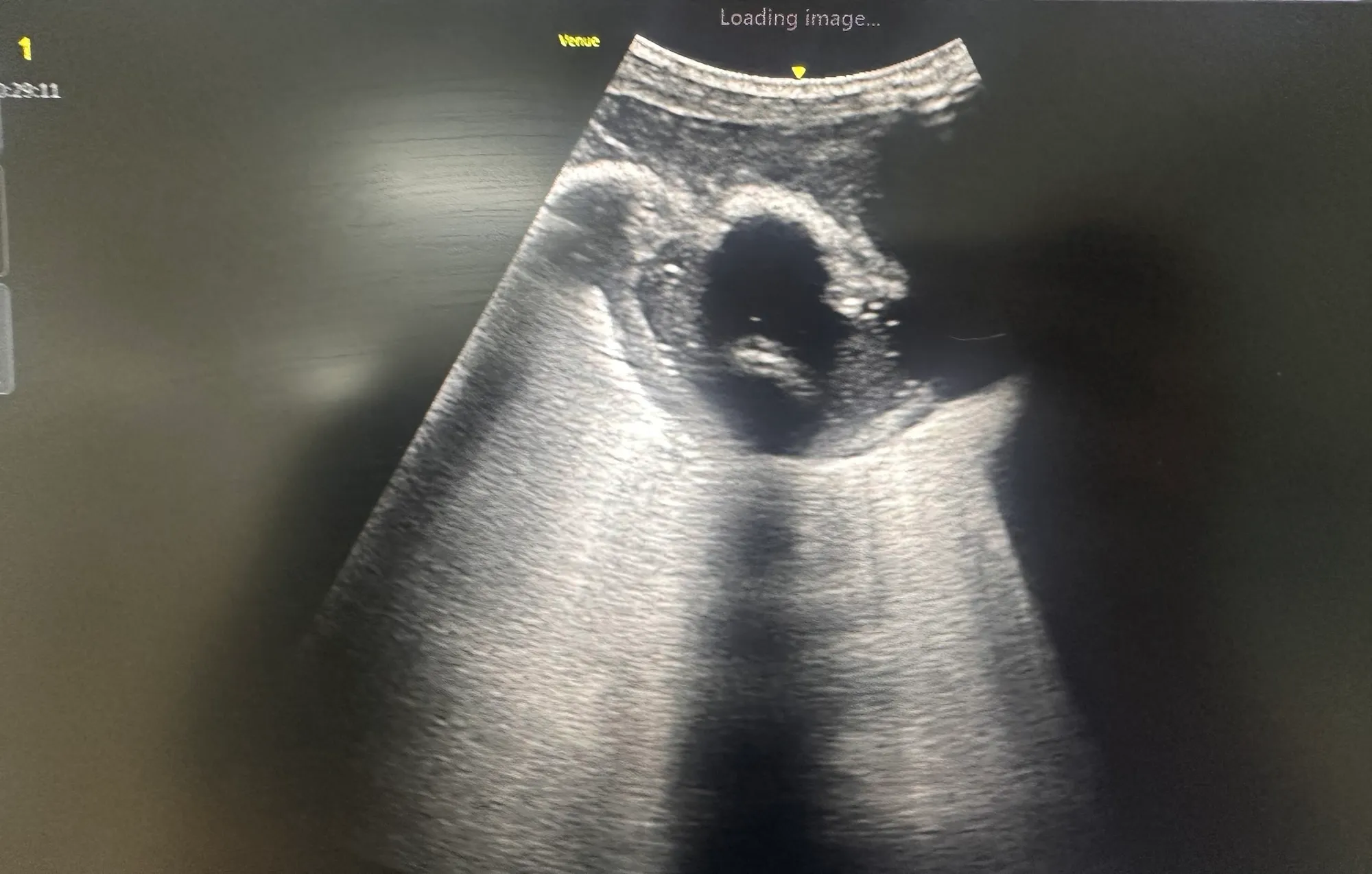
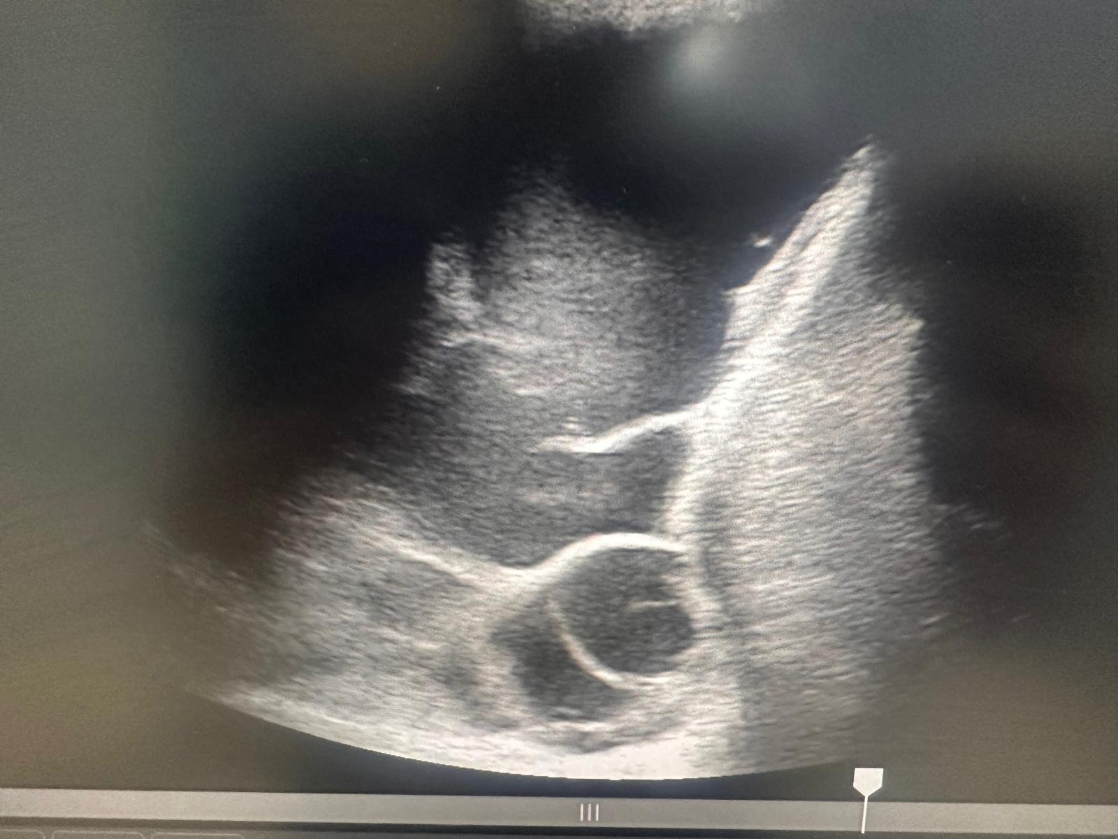
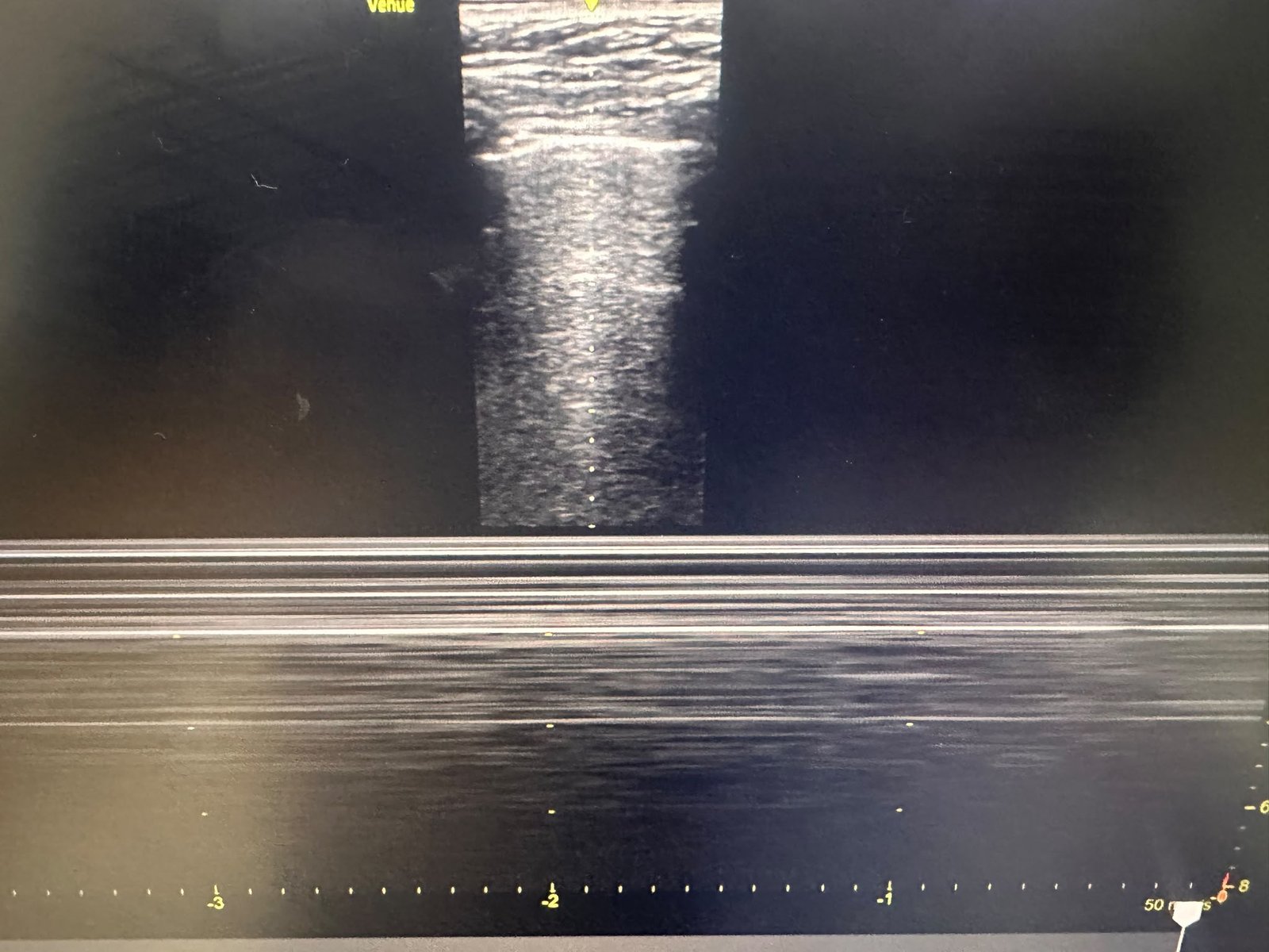
An 88-year-old woman presented with sepsis, worsening shortness of breath and a productive cough. Bedside lung POCUS demonstrated a complex, loculated Right sided pleural effusion containing internal septations,features concerning for a complicated parapneumonic effusion or empyema.
Pleural fluid sampling showed marked acidosis, supporting pleural infection. A chest drain was inserted under image guidance, allowing the infected fluid to be drained. Following drainage and intravenous antibiotics, her symptoms improved significantly. The post-drainage chest X-ray demonstrated substantial resolution of the pleural collection, and a respiratory referral was made for continued specialist care.
What causes a complex pleural effusion?
A complex effusion contains septations, debris or echogenic material rather than appearing completely anechoic. Important causes include:
- Complicated parapneumonic effusion or empyema
- Malignancy
- Haemothorax
- Tuberculosis
- Previous inflammation, surgery or pleural intervention
The ultrasound appearance alone does not establish the cause. Clinical context, pleural-fluid analysis, microbiology and where appropriate cytology and CT imaging remain essential.
How is it managed?
Management depends on the underlying cause and severity. Suspected pleural infection requires prompt antibiotics, diagnostic aspiration and pleural-fluid testing. Frank pus or pleural-fluid pH ≤7.2 generally indicates the need for chest-drain insertion when it can be performed safely. Persistent loculated collections may require intrapleural tPA and DNase or surgical assessment if adequate drainage cannot be achieved.
This case demonstrates how bedside POCUS can rapidly identify a clinically important pleural collection, guide safe intervention and allow treatment to begin without delay.
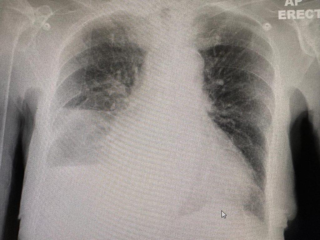
Pre Drainage CXR
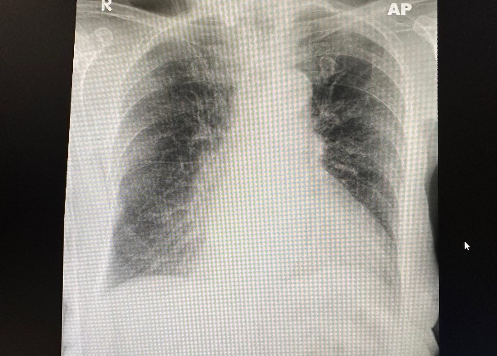
Post Drainage CXR