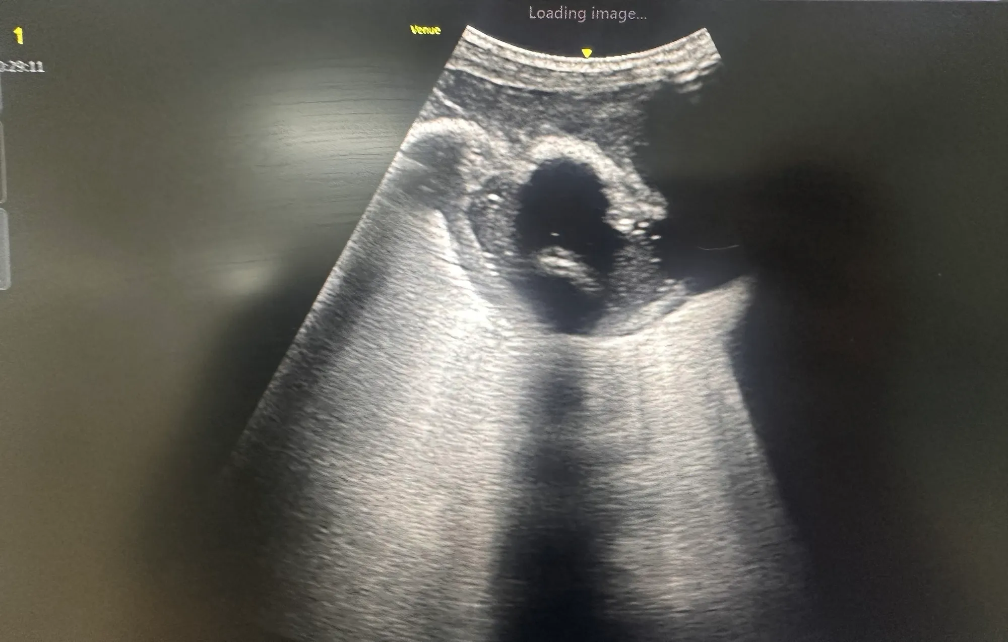
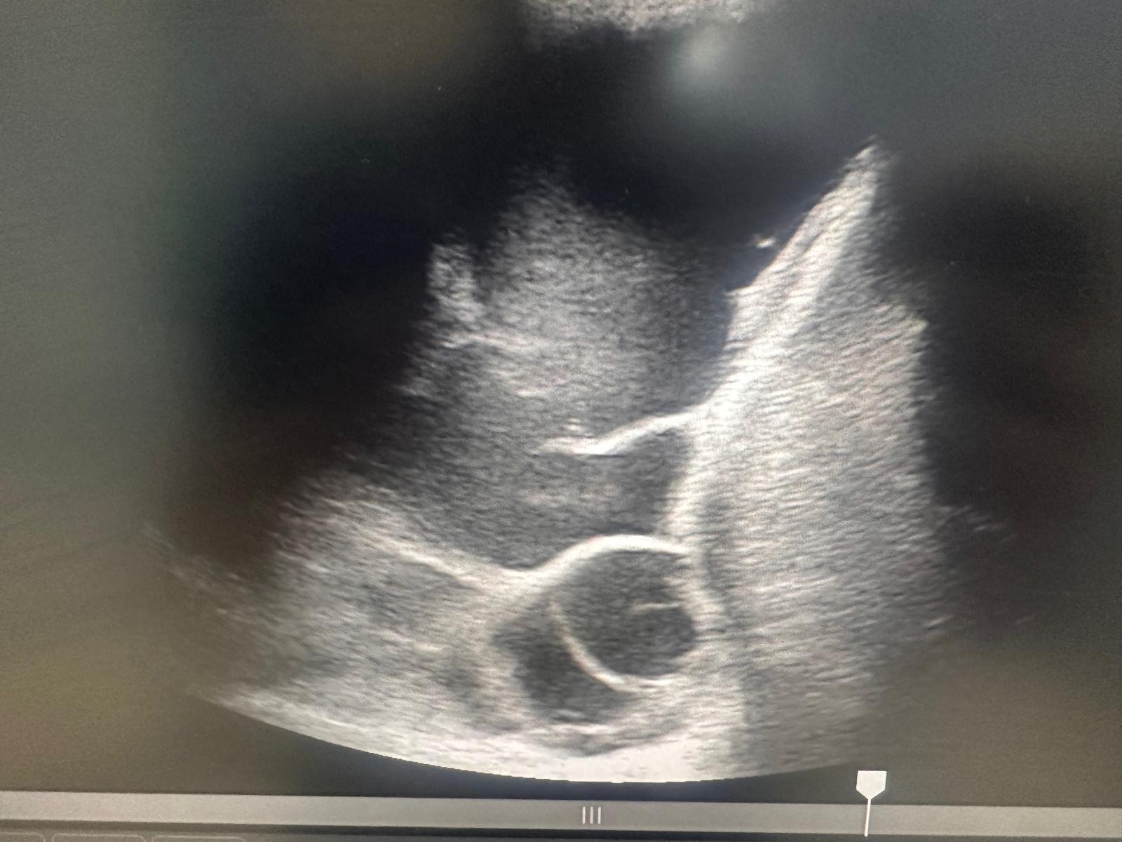
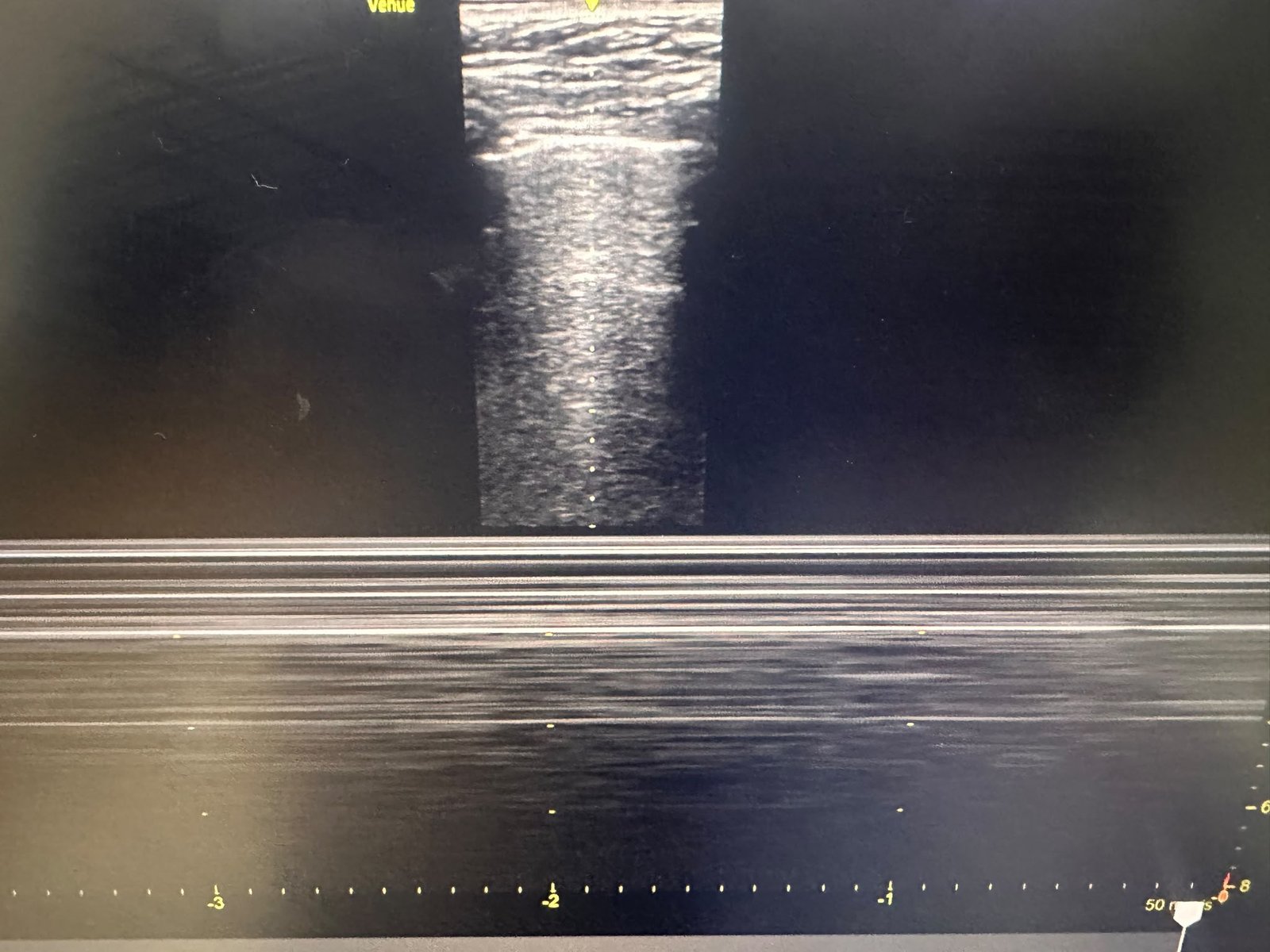
A 65-year-old man presented with severe shortness of breath, tachycardia and hypoxaemia requiring supplemental oxygen. Bedside cardiac POCUS demonstrated marked right ventricular dilatation in the parasternal short-axis view, raising immediate concern for acute right-heart strain and pulmonary embolism.
An urgent CT pulmonary angiogram subsequently confirmed pulmonary embolism. The diagnosis changed the direction of management, and anticoagulation with heparin was commenced.
POCUS Features of Right-Heart Strain
Findings that may support suspected pulmonary embolism include:
- Right ventricular dilatation
- An RV-to-LV diameter ratio greater than 1
- Interventricular septal flattening, producing a D-shaped left ventricle in the parasternal short-axis view
- Reduced right ventricular systolic function
- A plethoric inferior vena cava
- McConnell’s sign in some patients
Acute pulmonary embolism increases pulmonary vascular resistance, placing sudden pressure strain on the right ventricle. This can produce RV dilatation and septal flattening, which may be recognised immediately at the bedside.
These findings are not diagnostic of pulmonary embolism in isolation. Similar appearances may occur with chronic pulmonary hypertension, severe respiratory disease, right ventricular infarction or other causes of right-heart dysfunction. They must therefore be interpreted alongside the clinical presentation and confirmed with definitive imaging where appropriate.
This case demonstrates how cardiac POCUS can rapidly identify right-heart strain in a critically breathless patient, strengthen suspicion of pulmonary embolism and accelerate definitive investigation and treatment.