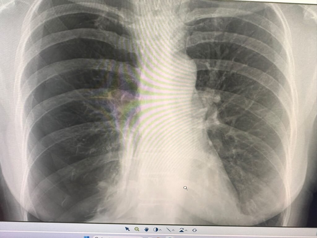
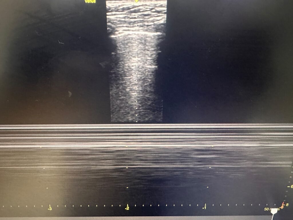
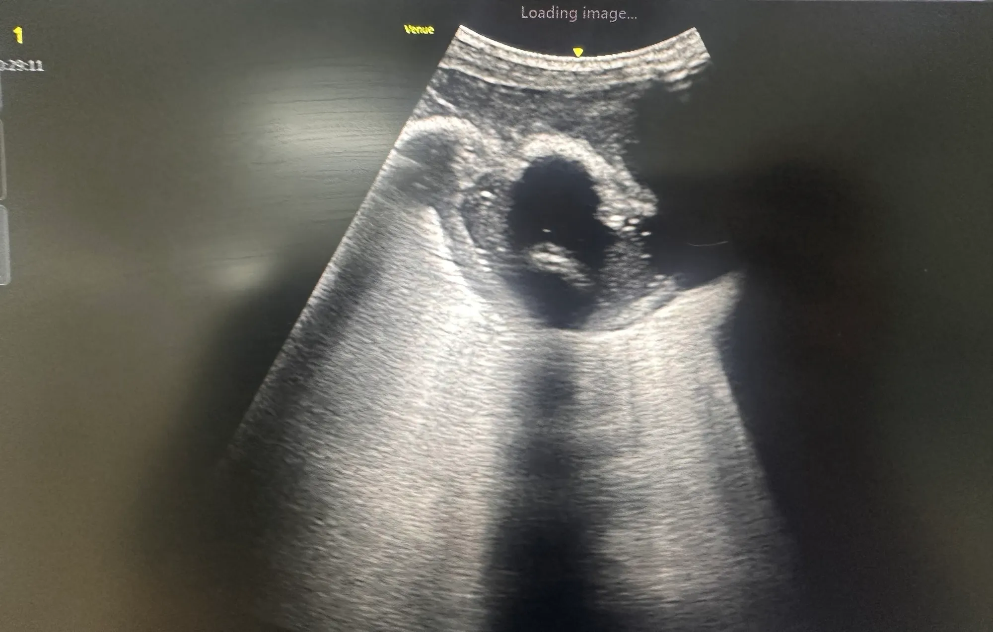
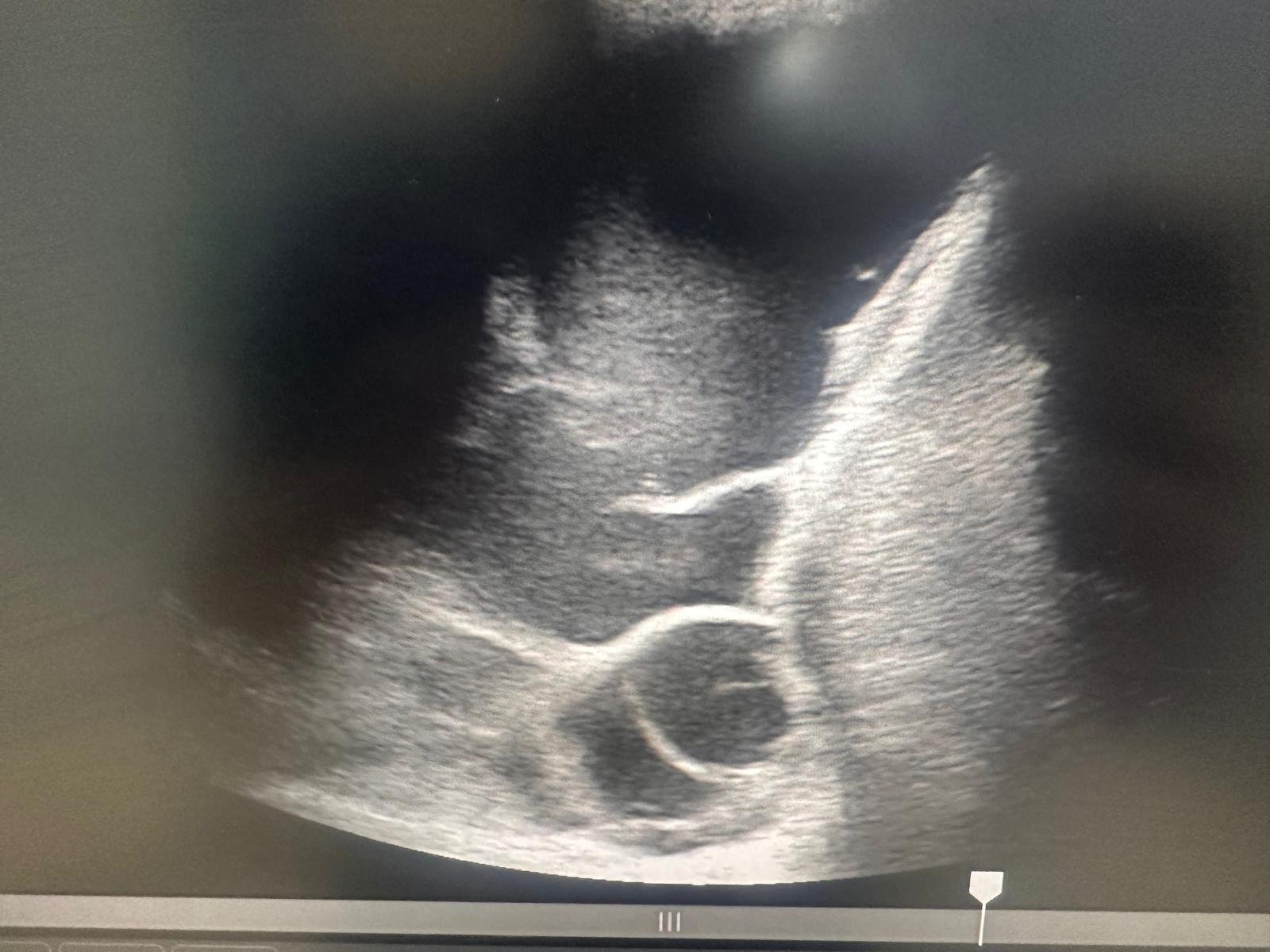
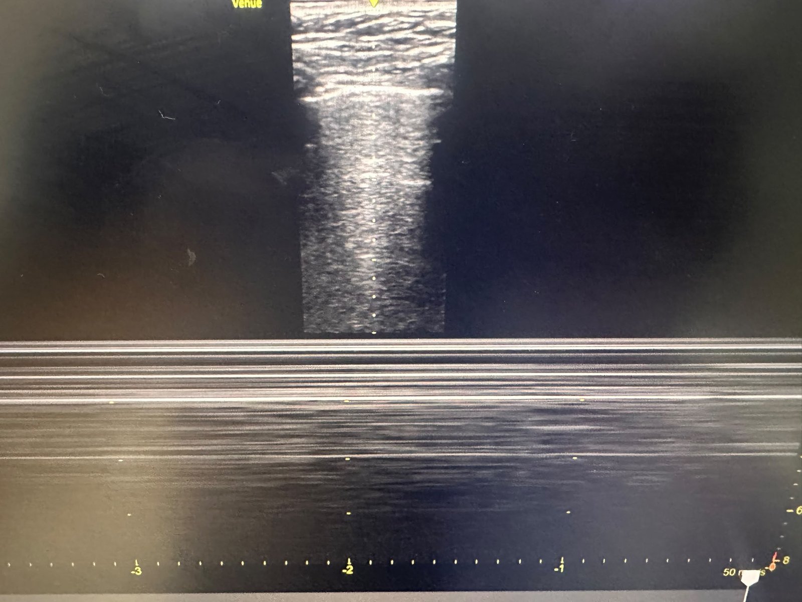
A 68-year-old woman presented with pleuritic chest pain and acute shortness of breath. Bedside lung POCUS demonstrated absent lung sliding and a clear lung point, strongly indicating a right-sided pneumothorax. A subsequent chest X-ray confirmed the diagnosis.
A chest drain was inserted, resulting in marked improvement in her symptoms. Follow-up imaging demonstrated successful re-expansion of the lung and resolution of the pneumothorax.
Key ultrasound findings
Important POCUS features of pneumothorax include:
- Absent lung sliding
- Absent B-lines
- Barcode or stratosphere sign on M-mode
- Lung point—the transition between sliding and non-sliding pleura
Absent lung sliding alone is not diagnostic and can occur in several other conditions, including apnoea, pleural adhesions, severe bullous disease and following pleurodesis. However, identifying a lung point in the appropriate clinical setting is highly specific for pneumothorax.
This case demonstrates the value of incorporating lung POCUS into the immediate assessment of acute breathlessness. It can provide a rapid bedside diagnosis, support prompt treatment and reduce delays while awaiting conventional imaging.